Radiation Survey Meter Selection Guide

Radiation safety programs often refer to handheld instruments simply as "survey meters," but that label hides an important truth: not all survey meters are designed to answer the same question. The right instrument depends on whether you are hunting for surface contamination, measuring an exposure rate, confirming x-ray leakage, or identifying an isotope, and choosing the wrong one leads to confusing data and inefficient response.
Some instruments are best for finding surface contamination. Others are better for measuring leakage or scatter around x-ray equipment. Some are optimized for low-level detection, while others are intended for more quantitative exposure-rate assessment. In medical physics and nuclear medicine, choosing the wrong instrument can lead to confusion, poor measurements, or inefficient response during routine surveys and contamination events.
That is why understanding how each detector works matters just as much as knowing how to operate it. This article compares five representative instruments, the technical principles behind them, and the survey question each one is built to answer.
Introduction
A radiation survey meter is a handheld instrument that detects and quantifies ionizing radiation so that physicists, technologists, and radiation safety officers can verify safe conditions. The category spans several distinct detector physics, and each is optimized for a different task. The goal of this guide is practical: help facilities in nuclear medicine and diagnostic imaging match the detector to the measurement, rather than treating every "survey meter" as interchangeable.
For Diagnostic Radiation Physics Services (DRPS) clients across Florida, Maryland, Virginia, Washington DC, California, and Nevada, instrument selection is a recurring theme in radiation safety program reviews. The instruments below are common in U.S. medical facilities, and the selection logic generalizes to whatever comparable models a department already owns.
Why Detector Type Matters
The detector type matters because the survey question dictates the physics you need. When a physicist, technologist, or radiation safety officer (RSO) performs a radiation survey, the real question is usually one of the following:
- Is contamination present?
- Where is the hotspot?
- What is the actual dose rate?
- Is this leakage or scatter within expectations?
Those are not the same question, and no single meter is ideal for every situation. A strong radiation safety program uses the appropriate detector for the task, supports staff training, and avoids treating all survey meters as interchangeable tools.
Below are five common examples relevant to medical physics and nuclear medicine workflows: a GM contamination meter, a hybrid survey meter, a specialized x-ray survey sensor, a pressurized ion chamber, and an imaging survey meter.
Ludlum GM Meter
The Ludlum GM meter is the classic contamination survey instrument, and it answers one question well: "Is there contamination here, and where is it?"
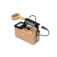
A classic example of a contamination survey instrument is a Ludlum Model 3 paired with a GM pancake probe (such as the Model 44-9). In many nuclear medicine departments, this is one of the most familiar survey meter configurations1.
A Geiger-Mueller detector is a gas-filled tube operated in the Geiger region, typically at 850-1000 volts DC, with 900 volts being the nominal operating voltage for most halogen-quenched GM tubes2, 3. The tube is filled with an inert gas (usually neon or argon) and a halogen quenching agent (such as bromine or chlorine). When radiation enters the detector, it ionizes the gas and produces a cascade avalanche that propagates throughout the entire tube volume, generating a large uniform pulse that is easy to detect electronically4.
The pancake probe design features an ultra-thin mica window with an areal density of 1.4-2.0 mg/cm² and an effective diameter of approximately 44.5 mm (1.75 inches), providing a sensitive area of about 15 cm²5, 6. This thin window allows detection of low-energy beta particles (>60-100 keV) that would otherwise be absorbed by thicker detector materials. Alpha particles with energies exceeding approximately 3.5 MeV can also penetrate the window, though air gap between source and window significantly affects alpha detection efficiency5, 7.
Technical Characteristics of GM Detection
The GM detector operates in the Geiger plateau region, where output pulse height becomes independent of incident radiation energy and type. The plateau typically extends over approximately 150-200 volts with a slope of 6-10% per 100 volts3, 8. Operating voltage is set 25-50 volts above the plateau "knee" to ensure stable performance2.
A critical limitation is dead time, the recovery period after each discharge during which the detector cannot register new events. For halogen-quenched GM tubes, dead time typically ranges from 20-100 microseconds, depending on operating voltage9, 10, 11. At the lower end of the operating voltage range, dead times can be as short as 9-26 μs, but increase to 250-300 μs at higher voltages due to charge multiplication effects9. This limits accurate counting to approximately 10³ counts per second; at higher count rates, significant dead time correction is required11.
The Ludlum Model 3 features adjustable high voltage from 400 to 1500 volts DC, a fixed threshold of 40 mV ± 10 mV, and selectable response times of either fast (4 seconds) or slow (22 seconds) from 10% to 90% of final reading2, 12. Operating range is typically 0-200 mR/hr or 0-500,000 counts per minute (cpm) using four-decade range selection (×0.1, ×1, ×10, ×100)1, 12.
What It Is Good For
- surface contamination surveys
- package surveys
- hot lab bench and floor checks
- identifying localized hotspots
- routine contamination control rounds
- alpha detection (when source-to-window air gap is minimized)
Advantages
- rugged and familiar design with >2000 hour battery life
- fast response to radiation presence
- effective for contamination finding due to large sensitive area
- pancake probes work well for close surface surveys
- halogen quenching provides long tube lifetime (unlike organic-quenched tubes which degrade over time)13
- high sensitivity: typical gamma sensitivity of 2,500-6,000 cpm/mR/hr for ¹³⁷Cs5, 14
Limitations
- not ideal for accurate dose-rate measurement (pulse height independent of energy)
- limited energy discrimination, cannot distinguish isotopes
- response depends on probe geometry and window characteristics
- susceptible to the fold-back effect: very high radiation fields can continuously retrigger the tube during recovery, producing pulses too small to count and falsely indicating low levels11
- dead time losses at high count rates (>10⁴ cpm) require correction
- mica window is fragile and can be damaged by physical contact
From a nuclear medicine perspective, a GM meter is often the right tool when the question is: "Is there contamination here, and where is it?"
RaySafe 452 Survey Meter
The RaySafe 452 is a hybrid survey meter that combines silicon diodes and a GM detector to handle both low-level contamination and quantitative dose-rate work in one instrument.
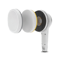
The RaySafe 452 takes a different approach from a traditional single-detector meter. It is a hybrid survey meter that combines multiple detector technologies into one instrument: a silicon sensor cluster (semiconductor diodes) and a thin-walled energy-compensated Geiger-Müller pancake detector (15.55 cm² sensitive area)15, 16.
This matters because different detector types bring different strengths. Semiconductor silicon diodes generate charge carriers directly in the detector material when radiation interacts with it, providing fast response and accuracy at higher dose rates. The silicon sensor cluster enables excellent performance in the diagnostic x-ray energy range (20 keV to 2 MeV per IEC 60846-1)16. The thin-walled GM tube provides high sensitivity and fast response time (~2 seconds to detect a step from 0.2 to 2 μSv/h) even at very low dose rates15, 16.
Technical Architecture and Performance
The RaySafe 452 uses interchangeable lids to switch measurement modes: one lid configuration for air kerma (Gy or R) and ambient dose equivalent (Sv or rem) measurements, and another for counts (cps or cpm) with alpha and beta detection capability15, 16. This design provides a wide and flat energy response across gamma, x-ray, beta (>50 keV), and alpha (>4 MeV) radiation17.
Key specifications include15, 16, 17:
- Energy range: 10 keV (gamma/x-ray), 50 keV (beta), 4 MeV (alpha)
- GM detector specifications:
- Sensitive area: 15.55 cm² behind 79% open steel grid
- Range: 0 to 20,000 cps (0 to 1.2 Mcpm)
- Automatic dead time correction (linearity within -10% to +30%)
- Typical gamma sensitivity: 6 cps/μGy/h (3,000 cpm/mR/h) for ¹³⁷Cs
- Typical background: 0.5 cps (30 cpm) at 0.1 μSv/h
- Dose rate range: 0 μGy/h to 1 Gy/h, 0 μSv/h to 1 Sv/h
- Minimum linac frequency: 100 Hz at T < 30°C
- Compliance: IEC 60846-1
- Environmental rating: IP 64 (dust proof and water resistant)
The instrument features automatic data storage (dose rate saved every second), PC software connectivity via USB, and alarm threshold settings15, 16. The combination of silicon diodes for higher dose rates and the GM detector for low-level sensitivity creates a versatile instrument with broad dynamic range.
What It Is Good For
- mixed radiation survey tasks spanning multiple applications
- x-ray tube leakage and wall leakage measurements
- scattered room radiation assessment
- contamination surveys (with GM detector mode)
- environmental radiation monitoring
- non-destructive testing applications
- departments that want one versatile general-purpose meter
- workflows spanning x-ray and radioactive material handling
Advantages
- broader capability than single-technology instruments
- supports multiple survey applications in one device
- modern digital interface with automatic data logging
- useful for departments with varied radiation tasks
- no manual corrections or settings required for different energies
- flat energy response reduces measurement uncertainty
- compliant with international standards (IEC 60846-1)
Limitations
- more complex than a basic GM meter
- users still need to understand which detector response is most relevant for the task
- higher cost than simple contamination meters
- may not outperform specialized instruments in every niche application
- requires understanding of interchangeable lid configurations
For many facilities, the RaySafe 452 is attractive because it functions as a generalist meter. It is particularly useful in departments that do not want separate handheld meters for every workflow. The hybrid detector approach provides both the sensitivity needed for contamination work and the accuracy required for dose rate measurements around imaging equipment.
RaySafe X2 Survey Sensor
The RaySafe X2 Survey Sensor is a specialized diagnostic x-ray instrument: it answers "What is the leakage or scatter around this x-ray system?" and is not intended for nuclear medicine contamination control.
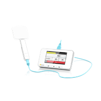
The RaySafe X2 Survey Sensor is more specialized. It is primarily intended for diagnostic x-ray leakage and scatter measurements, not routine nuclear medicine contamination control.
The detector is based on an energy-compensated silicon diode array18. A semiconductor detector works by generating electron-hole pairs directly in the silicon material when radiation interacts with it. When a photon deposits energy in the depleted silicon region, the number of charge carriers produced is proportional to the photon energy. Compared with gas-filled detectors, silicon's approximately 2,000 times higher density than air, combined with approximately 10 times lower energy per charge pair production (3.6 eV vs 34 eV in air), results in approximately 18,000 times higher signal per volume19. This enables construction of compact detectors with fast response.
Energy Compensation Mechanism
A fundamental challenge with silicon diode detectors is their strong energy dependence, the photoelectric effect cross-section in silicon (Z=14) varies dramatically with photon energy, particularly below 100 keV. Silicon diodes have inherently higher sensitivity to low-energy photons due to increased photoelectric absorption probability19, 20. This would cause the detector to over-respond at lower energies if left uncompensated.
Energy compensation is achieved by surrounding the silicon sensor with a high-Z material filter (such as tin, copper, or tungsten) of carefully selected thickness and design19, 20. The filter preferentially absorbs low-energy photons, flattening the overall energy response to approximate ambient dose equivalent H*(10) or air kerma across the diagnostic x-ray energy range. This allows a single calibration factor to be used across a wide energy spectrum18, 20.
The X2 Survey Sensor provides the unique capability to switch energy response between Air Kerma (Gy or R) and Ambient Dose Equivalent (Sv), effectively functioning as several instruments in one18. While ambient dose response is virtually flat in the x-ray range, this flexibility makes it suitable for various medical physics applications.
Technical Advantages for Diagnostic X-ray Work
Unlike pressurized ion chambers, silicon-based sensors can be shipped via air or ground transportation without special considerations or regulatory restrictions (no pressurized vessel)18. The sensor displays dose, dose rate, mean energy, and time, along with a dose rate waveform visualization. A real-time dose rate bar and audible "ticker" proportional to dose rate provide immediate feedback during surveys18.
The sensor offers two trigger modes, manual and automatic, making it excellent for low dose rate measurements even in the primary beam of x-ray machines18. This versatility extends its utility beyond simple leakage surveys to include comprehensive equipment performance assessment.
What It Is Good For
- diagnostic x-ray leakage surveys (tube housing, image intensifier)
- scatter measurements around radiographic and fluoroscopic systems
- room barrier shielding verification
- x-ray equipment performance assessment and quality control
- medical physics testing in diagnostic imaging environments
- primary beam measurements at low dose rates
Advantages
- optimized for diagnostic x-ray energy range with flat energy response
- compact, lightweight design
- well suited for leakage and scatter measurements
- integrates with RaySafe X2 platform ecosystem
- no shipping restrictions (unlike pressurized ion chambers)
- displays mean energy, useful for beam quality assessment
- switchable energy response (air kerma vs ambient dose equivalent)
Limitations
- not intended for routine nuclear medicine contamination surveys
- not the right choice for spill follow-up on surfaces
- more specialized than general-purpose survey instruments
- limited to photon detection (no beta or alpha capability)
- silicon detector subject to radiation damage at very high cumulative doses
This is an excellent tool when the question is: "What is the leakage or scatter around this x-ray system?" It is not the right instrument when the task is to find removable contamination on a hot lab countertop or floor. For a closely related look at how diagnostic imaging equipment is validated, see our guide on CT protocol optimization and dose-image quality balance.
Fluke Biomedical Ion Chamber Meter
The Fluke Biomedical ion chamber meter is the quantitative workhorse for leakage, scatter, and shielding surveys: it answers "How much radiation field is present here?"
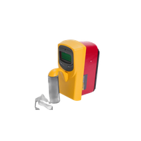
A widely used example of an ion chamber survey meter is the Fluke Biomedical 451P or the related 451B family. These instruments are standard tools for leakage and scatter surveys around x-ray equipment and radiation therapy areas.
An ion chamber works fundamentally differently from a GM detector. Instead of producing a large avalanche pulse that saturates the entire detector volume, radiation creates ion pairs in the gas inside the chamber (typically air or an air-equivalent gas mixture), and the instrument measures the current produced by collecting those ions under an applied electric field. The chamber operates in the ionization region, where the number of ion pairs collected is directly proportional to the energy deposited by radiation21.
Pressurized Ion Chamber Technology
The 451P uses a 230 cm³ pressurized ionization chamber filled to 8 atmospheres (125 psi)21, 22. Pressurization serves multiple purposes:
-
Enhanced sensitivity: Eight times atmospheric pressure provides eight times more gas molecules, dramatically increasing the number of ion pairs produced per unit radiation exposure. This enables μR/hr resolution, critical for measuring low-level leakage21, 22.
-
Improved energy response: The pressurized air-equivalent gas mixture provides a more uniform response across the x-ray and gamma energy spectrum (from 25 keV and above)22.
-
Better dose rate linearity: Ion chambers exhibit excellent linearity over a wide dose rate range because they operate in the ionization region where recombination effects are minimal at typical survey-level exposure rates.
The 451P specifications include21, 22:
- Operating ranges: 0-500 μR/h, 0-5 mR/h, 0-50 mR/h, 0-500 mR/h, 0-5 R/h (or equivalent in Sv/h)
- Accuracy: ±10% of reading between 10% and 100% of full scale
- Response time: 1.8 seconds for most ranges
- Energy detection: Beta above 1 MeV; gamma and x-rays above 25 keV
- Calibration: Factory calibrated to ¹³⁷Cs standard
The instrument features auto-ranging, auto-zeroing, automatic backlight, and can simultaneously display radiation rate and accumulated dose. A "Freeze Mode" records peak rate during surveys21. RS-232 communications interface enables data logging via optional Windows-based software21.
Ion Chamber Measurement Principles
Unlike pulse-mode detectors (GM and scintillators), ion chambers operate in current mode. The continuous collection of ionization current provides inherent averaging, making ion chambers less susceptible to statistical fluctuations than counting detectors. This is particularly valuable when measuring steady-state fields around x-ray rooms or radiation therapy vaults.
The chamber's air-equivalent response means measurements correlate well with exposure and dose quantities. Tissue-equivalent chambers can be used when dose-to-tissue assessment is the primary goal, though air-equivalent chambers remain standard for most medical physics surveys.
What It Is Good For
- x-ray leakage measurements (tube housing, barriers)
- shielding surveys and barrier adequacy verification
- room exposure-rate assessments
- scatter measurements around diagnostic and therapeutic equipment
- higher-field quantitative surveys
- radiation therapy area surveys (treatment rooms, mazes)
- occupational area monitoring
Advantages
- better for true dose-rate or exposure-rate measurement (proportional response)
- appropriate for leakage and shielding work
- stable and well suited to room surveys over extended periods
- standard choice for many medical physics regulatory compliance applications
- excellent linearity and energy response
- minimal dead time effects (current mode, not pulse counting)
- widely recognized by regulatory agencies
Limitations
- less sensitive than a GM pancake probe for finding tiny contamination spots
- not ideal for detailed surface contamination searches
- bulkier and less convenient for close-geometry contamination mapping
- pressurized chamber may require special shipping considerations
- typically more expensive than basic GM contamination meters
- slower response when searching for small localized hotspots
An ion chamber is often the right tool when the question is: "How much radiation field is present here?" rather than "Where is the small contaminated spot?" Ion chamber surveys underpin shielding adequacy work, which is discussed further in our lead shielding design principles guide.
RAVIN CAM Imaging Survey Meter
The RAVIN CAM is an imaging survey meter: it overlays a real-time radiation heat map on optical video and identifies isotopes by spectroscopy, answering "Exactly where is the radiation, what isotope is it, and how is it distributed?"
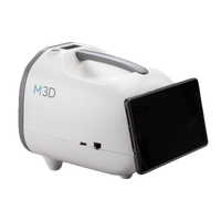
The RAVIN CAM represents a newer category: the imaging survey meter. Rather than only giving the user a numerical reading or audible count rate, the RAVIN CAM provides real-time visualization of radiation distribution by overlaying a radiation heat map on optical images or live video streams23, 24.
According to M3D, the RAVIN CAM uses a >19 cm³ pixelated cadmium zinc telluride (CZT) detector, a room-temperature solid-state semiconductor detector with exceptional energy resolution and imaging capability23, 24. The system provides a 4π radiation field of view, meaning it can detect radiation from all directions simultaneously, not just from a narrow cone24, 25.
Pixelated CZT Detector Technology
Cadmium zinc telluride (Cd₁₋ₓZnₓTe, typically with x ≈ 0.1) is a high-Z (Z_Cd = 48, Z_Te = 52) semiconductor compound that operates at room temperature without cryogenic cooling26, 27. Unlike silicon detectors, CZT's higher atomic number and density provide much better stopping power for gamma rays in the medical imaging energy range (30-662 keV per RAVIN CAM specifications)23.
Pixelated detector architecture divides the detector volume into an array of individual pixels, each functioning as an independent spectrometer. When a gamma ray interacts in the detector, the position of interaction can be determined by identifying which pixel(s) collected the charge. Modern pixelated CZT detectors achieve energy resolutions of 1-2% FWHM at 662 keV26, 27, dramatically better than NaI scintillators (~7-8% FWHM) and approaching high-purity germanium performance without requiring liquid nitrogen cooling.
The RAVIN CAM specifications indicate23, 24:
- Detector: >19 cm³ pixelated CZT
- Imaging/spectroscopy range: 30 to 662 keV
- Energy resolution: 1.1% FWHM at 662 keV (approximately 7 keV)
- Minimum activity for imaging: 37 kBq (1 μCi) at 1 meter for 140 keV photons
- Dose rate range: starting at 1 nSv/hr
- Dose rate accuracy: ±10% at 662 keV
- Dimensions: H 22 cm × W 20 cm × L 25.5 cm
- Weight: 2.3 kg
This exceptional 1.1% FWHM energy resolution enables isotope identification through gamma spectroscopy, the detector can distinguish photopeaks from different radionuclides and identify mixed contamination sources23, 24.
Imaging Capability and Operational Advantages
The key innovation is spatial imaging combined with spectroscopy. Traditional survey meters tell you radiation is present and how much is detected at a point. The RAVIN CAM shows where the radiation is located in three-dimensional space and what isotope is present.
M3D reports the device is 3 times faster at source localization compared to traditional GM probes24. Instead of slowly scanning a room point-by-point while watching count rate fluctuations, the user can sweep the area while viewing a live heat map overlay that immediately highlights contamination locations. This significantly reduces survey time and radiation exposure during contamination response.
The system captures high-resolution images and spectroscopy data simultaneously, supporting applications including24, 25:
- Contamination cleanup: Visualize spills, identify isotopes, verify decontamination effectiveness
- Shielding verification: Image radiation penetration through barriers, identify shielding gaps
- Isotope identification: Distinguish between different radionuclides in mixed waste or contamination
- Patient monitoring: Assess dose rates around patients after radiopharmaceutical administration
- Waste handling: Identify and segregate radioactive waste by isotope
What It Is Good For
- contamination source localization and mapping
- shielding verification and barrier adequacy testing
- isotope identification and waste characterization
- hotspot visualization in three-dimensional space
- patient or room radiation imaging support
- investigations where "where is it coming from?" matters as much as "how much is there?"
- training and communication (visual demonstration of radiation fields)
- complex contamination events requiring rapid source identification
Advantages
- provides spatial visualization of radiation distribution
- may significantly speed up contamination response and source localization (3× faster than GM scanning per manufacturer)
- useful for troubleshooting shielding concerns and identifying gaps
- isotope identification capability through high-resolution spectroscopy (1.1% FWHM)
- can improve communication and training because users can see source distribution
- supports advanced workflows beyond simple count-rate surveying
- 4π field of view detects radiation from all directions
- room-temperature operation (no cryogenic cooling required)
- records images and video for documentation and analysis
Limitations
- more advanced and higher cost than standard survey meters
- does not replace routine calibrated contamination and exposure-rate instruments for regulatory surveys
- requires workflow integration and user training
- value depends on whether department has use cases that benefit from imaging
- operating temperature range limited (15-25°C optimal) compared to rugged field instruments
- heavier and bulkier than traditional handheld meters (2.3 kg)
- minimum detectable activity higher than sensitive GM contamination meters for low-energy beta emitters
The RAVIN CAM is most compelling when the question is not just "Is there radiation here?" but "Exactly where is it, what isotope is it, and how is it distributed?" For departments with higher-complexity nuclear medicine or radiation safety workflows, particularly those dealing with spills, mixed waste, or frequent contamination events, that spatial and spectroscopic capability can provide meaningful operational value24, 25. Departments handling spills will also benefit from our overview of decontamination best practices in nuclear medicine.
Key Technical Principles
Five detector physics underlie the instruments above, and understanding them is the difference between collecting data and interpreting it correctly:
- Geiger-Mueller avalanche (pulse mode): A single ionizing event triggers a full-tube discharge, producing a large, energy-independent pulse. Excellent for detecting the presence of contamination, but pulse height carries no energy information, so a GM meter cannot identify isotopes or accurately report dose rate. Dead time (20-100 μs) limits accurate counting above ~10³ cps9, 10, 11.
- Ionization current mode: Collected ion-pair current is proportional to energy deposited, giving accurate, stable exposure-rate and dose-rate readings with minimal dead time. The basis for leakage and shielding surveys21.
- Silicon diode (semiconductor, photon): Charge carriers are generated directly in silicon, yielding fast response and high signal per volume (~18,000× air), but strong low-energy over-response requires a high-Z compensation filter to flatten the diagnostic x-ray energy response18, 19, 20.
- Pixelated CZT (semiconductor, imaging spectroscopy): A high-Z, room-temperature detector array that localizes interactions per pixel and resolves photopeaks (1.1% FWHM at 662 keV), enabling both spatial imaging and isotope identification23, 26, 27.
- Energy compensation vs. energy independence: GM and silicon detectors must be engineered or filtered to behave predictably across energies; ion chambers achieve near-proportional response inherently. Knowing which applies prevents misreading a number that the detector physics never intended to deliver.
Clinical Impact
Matching the detector to the question directly affects patient and staff safety, survey efficiency, and regulatory defensibility. In practice:
- Faster, lower-dose contamination response. Using a GM pancake probe to localize a spill, then a calibrated ion chamber to quantify the field, minimizes the time staff spend in the radiation field, a direct application of ALARA. Imaging survey meters can further compress localization time (reported 3× faster than GM scanning)24.
- Defensible leakage and shielding data. Ion chamber and energy-compensated silicon measurements produce exposure-rate and dose-rate values that regulators and accreditation bodies recognize for barrier adequacy verification, which protects adjacent occupied areas and supports occupancy-factor assumptions in shielding designs.
- Accurate isotope and patient-release decisions. Spectroscopic identification (CZT) supports waste segregation and post-administration patient dose-rate assessment, both of which feed compliance with NRC patient-release and waste-handling expectations under 10 CFR 35 and 10 CFR 20.
- Fewer measurement errors. A technologist who understands that a GM pulse height is energy-independent will not misuse it for dose-rate reporting, and one who recognizes dead time and the fold-back effect will not be fooled by a falsely low reading in a high field.
Practical Tips for Instrument Selection
Start from the question, not the catalog. The best meter is the one that matches what you are trying to measure:
- Define the survey question first. "Is contamination present and where?" points to a GM pancake probe. "How much field is here?" points to an ion chamber. "What is the leakage around this x-ray system?" points to an energy-compensated silicon sensor. "Where exactly, and what isotope?" points to an imaging survey meter.
- Match the detector to the radiation type and energy. Confirm the instrument responds to the particles and energies you expect (beta thresholds, alpha capability, low-energy photon response) before relying on it.
- Calibrate and check before use. Verify current calibration (typically to a ¹³⁷Cs standard), perform a daily battery and response check against a known check source, and confirm the operating voltage sits above the plateau knee for GM instruments.
- Respect dead time and the fold-back effect. At high count rates, apply dead time correction; in very high fields, cross-check a GM reading against an ion chamber to avoid a falsely low fold-back indication11.
- Plan for more than one instrument. Most medical physics programs need at least a GM contamination meter and an ion chamber; specialized or imaging instruments are added when workflow justifies them.
- Train staff on interpretation, not just operation. The most common survey errors come from using the right meter the wrong way, or the wrong meter for the task.
Practical Comparison
| Instrument | Detection principle | Best use | Main limitation |
|---|---|---|---|
| Ludlum GM meter with pancake probe | Gas-filled GM detector (halogen-quenched, ~900V, 20-100 μs dead time) | Contamination surveys and hotspot finding | Not ideal for accurate dose-rate measurement; dead time losses at high count rates |
| RaySafe 452 | Hybrid detector: silicon diode cluster + energy-compensated GM pancake (15.55 cm²) | Broad mixed-use surveying across x-ray and nuclear medicine | More complex and costlier than simple GM meter; requires understanding of detector modes |
| RaySafe X2 Survey Sensor | Energy-compensated silicon diode array | Diagnostic x-ray leakage and scatter (20 keV - 2 MeV) | Not a contamination survey tool; limited to photon detection |
| Fluke 451P / 451B ion chamber | 230 cm³ pressurized air ionization chamber (8 atmospheres) | Quantitative leakage, scatter, shielding, and room surveys | Less sensitive for tiny contamination spots; bulkier than pancake probes |
| RAVIN CAM | >19 cm³ pixelated CZT detector (1.1% FWHM energy resolution, 30-662 keV) | Radiation visualization, contamination localization, isotope identification, shielding verification | Higher complexity and cost; operating temperature limitations |
Regulatory Considerations
Survey instrument selection and use are bounded by federal and state radiation regulations, and proper instruments are a prerequisite for compliance, not just good practice. Key references include:
- NRC and Agreement State rules: Survey requirements for licensed material flow from 10 CFR 20 (Standards for Protection Against Radiation) and 10 CFR 35 (Medical Use of Byproduct Material), which require area surveys, contamination surveys, and instruments appropriate to the radiation being measured. In NRC Agreement States, an equivalent state program applies, for example, Florida 64E-5 administered by the Florida Bureau of Radiation Control governs facilities in Florida. DRPS also supports clients in Maryland, Virginia, Washington DC, California, and Nevada, each operating under its own NRC or Agreement State framework.
- Calibration: Survey meters used for regulatory surveys must be calibrated, typically annually, with the calibration source and method documented. Most medical instruments are calibrated to a ¹³⁷Cs standard.
- Shielding design and verification: Barrier adequacy surveys with ion chamber or energy-compensated instruments support shielding designs prepared under NCRP Report No. 151 for diagnostic and dental imaging facilities.
- International instrument standards: Hybrid and silicon instruments referenced here comply with IEC 60846-1 for ambient and directional dose-equivalent rate meters.
Always confirm the current, jurisdiction-specific requirement, the items above are accurate at a general level but state implementations vary. For a deeper look at where survey-related compliance commonly breaks down, see common radiation safety violations and how to avoid them and the Florida radiation safety requirements for imaging centers.
Frequently Asked Questions (FAQs)
What is the difference between a GM survey meter and an ion chamber?
A Geiger-Mueller (GM) meter counts individual ionization events as large, energy-independent pulses, making it highly sensitive for finding contamination but poor for accurate dose-rate measurement. An ion chamber operates in current mode and measures the ionization charge proportional to energy deposited, giving accurate exposure-rate and dose-rate readings for leakage and shielding surveys.
Which radiation survey meter is best for nuclear medicine contamination surveys?
A GM pancake probe paired with a rate meter, such as a Ludlum Model 3 with a Model 44-9 pancake, is the standard choice. Its thin mica window and roughly 15 cm² sensitive area detect low-energy beta and some alpha particles, making it ideal for surface contamination, package surveys, and hotspot identification.
Why do ion chambers used for x-ray leakage surveys need to be pressurized?
Pressurizing the chamber, for example to 8 atmospheres in the Fluke 451P, packs about eight times more gas molecules into the same volume, producing more ion pairs per unit exposure. This boosts sensitivity to the μR/hr range needed to confirm leakage and shielding adequacy while preserving good energy response and dose-rate linearity.
Can a single survey meter handle every medical physics task?
No. Contamination detection, dose-rate measurement, x-ray leakage, and isotope identification are different questions that favor different detectors. A well-equipped radiation safety program typically combines a GM contamination meter, an ion chamber, and, depending on workflow, a specialized x-ray sensor or imaging survey meter.
What is an imaging survey meter and when is it worth using?
An imaging survey meter, such as the RAVIN CAM, overlays a radiation heat map onto live optical video and uses a pixelated CZT detector to identify isotopes by gamma spectroscopy. It is most valuable for rapid source localization, shielding gap detection, and mixed-contamination events, but it supplements rather than replaces calibrated meters for routine regulatory surveys.
Key Takeaways
- No single meter fits every survey. GM detectors find contamination, ion chambers quantify fields, silicon sensors handle x-ray leakage, and imaging meters localize and identify isotopes, the right tool depends on the question being asked.
- GM detectors are sensitive but energy-blind. A halogen-quenched GM tube (~900 V) produces an energy-independent pulse with 20-100 μs dead time, so it cannot accurately measure dose rate or identify isotopes2, 9.
- Ion chambers are the quantitative standard. A pressurized 230 cm³ chamber at 8 atmospheres delivers μR/hr resolution with excellent linearity, making it the recognized choice for leakage and shielding surveys21, 22.
- Energy compensation makes silicon diodes usable for dosimetry. A high-Z filter flattens silicon's low-energy over-response so one calibration factor works across the diagnostic x-ray range18, 19, 20.
- Pixelated CZT adds spatial imaging plus spectroscopy. At 1.1% FWHM resolution and a 4π field of view, it can locate a source and identify the isotope, reportedly localizing 3× faster than GM scanning23, 24.
- Instrument selection is an ALARA tool. Matching detector to task minimizes time in radiation fields and supports accurate, defensible compliance measurements28, 29.
How DRPS Can Help
Diagnostic Radiation Physics Services (DRPS) helps imaging centers, hospitals, and nuclear medicine departments select, calibrate, and correctly apply radiation survey instruments as part of a complete radiation safety program. Our board-certified medical physicists (PhD, DABR) support facilities across Florida, Maryland, Virginia, Washington DC, California, and Nevada with instrument selection guidance, survey procedure development, RSO support, leakage and shielding surveys, and contamination control program reviews aligned with NRC, Agreement State (including Florida 64E-5), and accreditation requirements. If you are unsure whether your survey meters match your workflow, or you are building out a new nuclear medicine or imaging service, reach out to DRPS for a tailored assessment.
Conclusion
Radiation survey instruments are essential tools in both nuclear medicine and diagnostic imaging physics, but they are only as useful as the user's understanding of their strengths and limitations.
A GM meter is excellent for contamination finding. An ion chamber is better for quantitative field assessment. A specialized x-ray survey sensor is ideal for leakage and scatter work. A hybrid meter may help bridge multiple workflows. An imaging survey meter like the RAVIN CAM adds a different capability altogether: the ability to visualize radiation in space and identify radionuclides through spectroscopy.
Good radiation safety does not come from owning more devices. It comes from matching the right detector to the right task, training staff appropriately, and using the results in a way that supports ALARA (As Low As Reasonably Achievable), compliance, and efficient clinical operations.
The ALARA principle stands as the cornerstone of radiation protection, advocating for minimizing radiation exposure while ensuring adequate diagnostic or therapeutic outcomes28, 29. ALARA is predicated on the conservative assumption that any radiation dose, regardless of magnitude, carries potential risk, thus necessitating continuous optimization of protection through time, distance, and shielding28, 29. Effective instrument selection directly supports ALARA by enabling efficient surveys that minimize time spent in radiation fields, accurate measurements that inform shielding adequacy, and rapid contamination localization that reduces exposure during cleanup operations.
Technical understanding of detector physics, combined with appropriate instrument selection and training, transforms radiation safety from a compliance exercise into an effective protection program that serves both occupational workers and patients.
Related Resources
- CT protocol optimization
- Lead shielding design principles
- Nuclear medicine decontamination best practices
- Common radiation safety violations
- Florida radiation safety requirements
References
- Ludlum Measurements, Inc. (2024). Model 3 general purpose survey meter. ludlums.com
- Ludlum Measurements, Inc. Ludlum Model 3 survey meter operation manual. fieldenvironmental.com
- LND, Inc. (2022). 7185 Pancake mica window alpha-beta-gamma detector specifications. lndinc.com
- Caltech Physics Laboratory. The Geiger counter and counting statistics. cco.caltech.edu
- Canberra Industries. GM pancake detectors technical specifications. pascalchour.fr
- University of California, Berkeley Environment, Health & Safety. G-M pancake detectors. ehs.berkeley.edu
- Direct Scientific. HP-265 G-M pancake probe specifications. drct.com
- Science First. EN-01 Geiger tube manual and specifications. sciencefirst.com
- Alaabdin, A. M. Z., et al. (2020). Experimental evaluation of the deadtime phenomenon for GM detector: deadtime dependence on operating voltages. Journal of Instrumentation, 15(12). experts.illinois.edu
- Scribd. (2026). Dead time of a Geiger Muller counter analysis. ru.scribd.com
- Wikipedia. (2002). Geiger–Müller tube. en.wikipedia.org
- Ludlum Measurements, Inc. Model 3 specifications sheet. fieldenvironmental.com
- Health Physics Society. (2025). Why is the slope of the G-M plateau greater for halogen-quenched tubes compared to organic-quenched? hps.org
- Victoreen. (2024). Process Geiger-Mueller (GM) detector and preamplifier specifications. victoreen.com
- RPD, Inc. (2026). RaySafe 452 radiation survey meter product overview. rpdinc.com
- RaySafe. RaySafe 452 radiation survey meter datasheet. raysafe.com
- Zoro. RaySafe 452 radiation survey meter specifications. zoro.com
- RaySafe. X2 Survey Sensor product information. raysafe.com
- Nilsson, B., & Montelius, A. (2003). Modeling silicon diode dose response in radiotherapy fields. Lund University Department of Medical Radiation Physics. diva-portal.org
- Ding, G. X., et al. (1977). Silicon diode detectors used in radiological physics measurements. Medical Physics, 4(6), 494-498. PubMed
- Fluke Biomedical. 451P pressurized μR ion chamber survey meter specifications. flukebiomedical.com
- TSG X-Ray. Pressurized μR ion chamber survey meter technical literature. tsgxray.com
- Southern Scientific. (2024). RAVIN CAM radiation imaging catalog. southernscientific.co.uk
- Southern Scientific. (2025). A new advanced imaging survey meter for nuclear medicine. southernscientific.co.uk
- Jokerst Scientific. (2025). M3D products—RAVIN CAM specifications. jokerst-scientific.com
- Bolotnikov, A. E., et al. (2010). Pixellated Cd(Zn)Te high-energy X-ray instrument. Journal of Instrumentation, 5. PubMed
- Boucher, Y. A. Analysis of cadmium zinc telluride detector performance and imaging characterization. University of Michigan CZT Laboratory. cztlab.engin.umich.edu
- RamSoft. (2021). 11 ALARA principles to minimize radiation exposure. ramsoft.com
- Landauer. (2024). What does ALARA mean and why is it so important? landauer.com